Shai M. Rozen, M.D., F.A.C.S.
1801 Inwood Road
Dallas, TX 75390
Phone: (214) 645-2353
Monday–Friday: 8 a.m.–5 p.m.
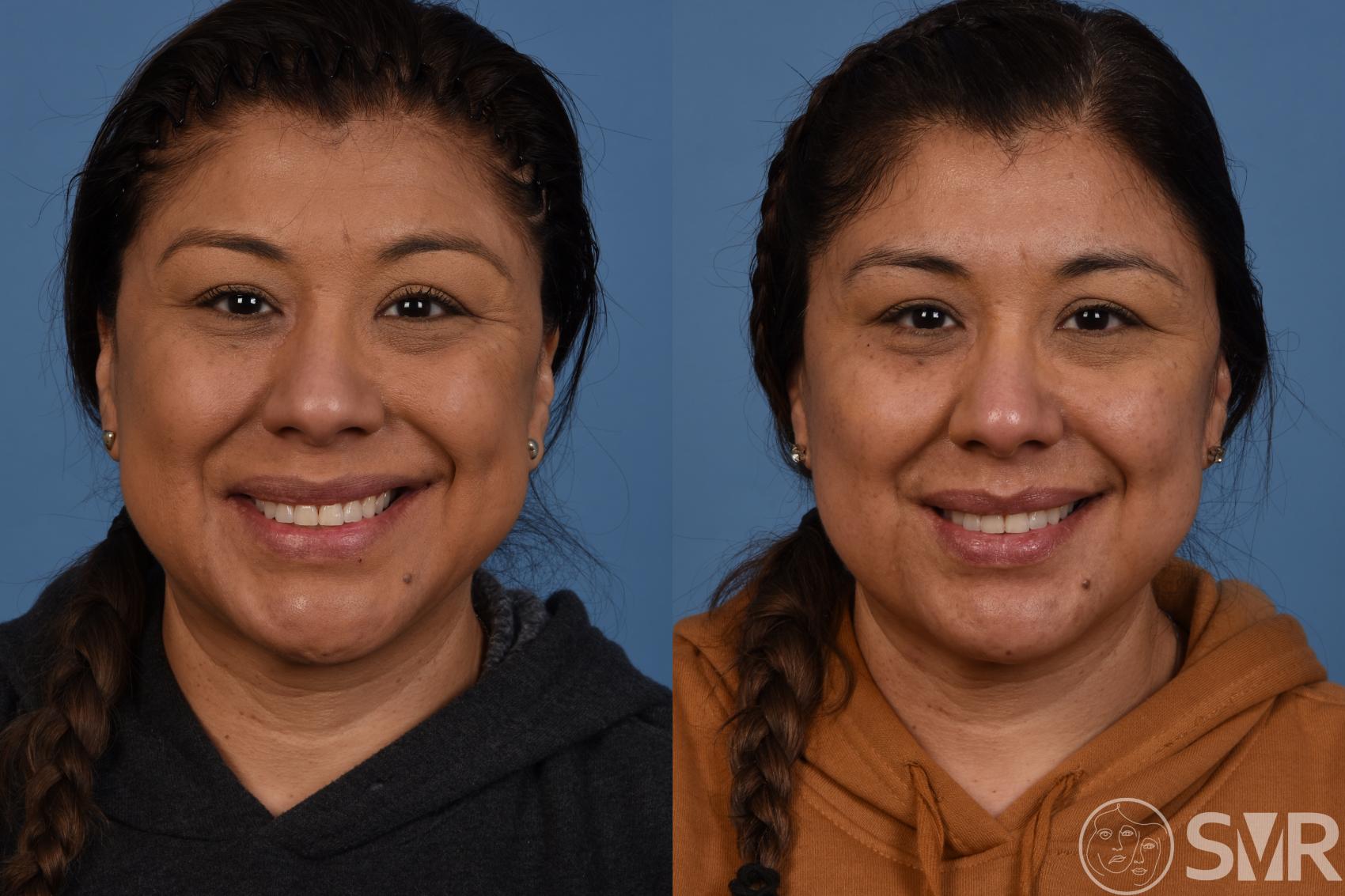
Selective Myectomies
Synkinesis can cause muscles around the eyes, mouth, and neck to become excessively tense or spastic or excessively weak, resulting in decreased functionality, facial asymmetry, and disfigurement. Myectomy is a surgical procedure that can help in both function and facial balance by weakening an excessively strong muscle on the synkinetic side and sometimes also by weakening a muscle on the healthy side that is not essential for function but may contribute to overall disfigurement and sometimes dysfunction.
Dr. Shai Rozen, a surgeon known nationally and internationally in the area of facial paralysis, and his team at the University of Texas Southwestern are very experienced in treating facial paralysis and synkinesis. Below is information on the use of selective myectomies in helping restore facial function, appearance, balance, and self-confidence for patients from across the U.S. and around the globe.
What is a myectomy?
Myectomy is the cutting of a muscle. Facial balance can be improved between the injured side and the healthy side of the face by performing selective myectomies on muscles in the synkinetic side, the healthy side, and often both sides depending on each patient’s unique presentation.

This figure depicts a myectomy of a muscle called the depressor anguli oris muscle that pulls down the corner of the mouth. The incision is performed within the mouth so there are no obvious scars on the skin.1
What happens to muscles in patients with synkinesis?
In patients with synkinesis, muscles in the injured side of the face may cause dysfunction and disfigurement in the following ways:
- Some muscles may be too strong or spastic (hypertonicity) and uncoordinated. Too much strength means that even at rest, these muscles create excessive pull and therefore disfigurement. Because they are uncoordinated, they activate and pull when they should not, also creating dysfunction.
- Some muscles are too weak. When the uninjured side of the face moves, the injured side cannot equally do so, thereby creating disfigurement and asymmetry.
Dr. Rozen provides a partial explanation for the seemingly paradoxical existence of stronger and weaker muscles adjacent to each other in his published research.2

This figure depicts the innervation of the depressor anguli oris and depressor labii inferioris muscles, partially explaining why adjacent muscle may be both strong and weak in synkinesis as described by Dr. Rozen.
Why are myectomies performed for patients with synkinesis?
It seems counterintuitive to cut a muscle when a patient has facial paralysis. But as explained above, muscles can be weak, atrophied (dead), or excessively strong. When a muscle is excessively strong and never rests (think of how your eyes tend to close up when the sun is very strong), it causes both disfigurement and asymmetry—and sometimes even discomfort or pain at the end of the day.
In other words, although these muscles are not atrophied, they are dysfunctional and function like a tight band that keeps pulling excessively, thereby disfiguring the face. A myectomy releases these muscles, similar to when we release a scar. By doing so, we help restore symmetry in 2 ways:
- Releasing the excessive pull of the muscle
- Stopping uncoordinated motion
Dr. Shai Rozen
Dr. Rozen is a board-certified plastic surgeon who co-created a facial paralysis specialty group with colleagues from otolaryngology & neurosurgery at the University of Texas Southwestern Medical Center.
Meet Dr. Rozen
Are myectomies also performed on the healthy side?
Though it also sounds counterintuitive to cut select muscles on the healthy side, there are several good reasons to assess the need for these myectomies. As mentioned previously, the synkinetic side suffers from a paradoxical problem of having some excessively strong (hypertonic) muscles that act like a scar and some weak muscles that can barely function.
In order to restore balance, it is often beneficial to weaken some muscles on the healthy side. This can sometimes be done with BOTOX® injections, but the results are temporary and many times not as accurate as surgery. For more permanent and accurate results, myectomy is often needed. By doing so, we can improve facial balance and symmetry without compromising function.
Which muscles are most commonly targeted for myectomies?
The most common and effective myectomies target muscles around the mouth.
Depressor Anguli Oris (DAO) Muscle
This muscle of the lower lip tends to be very strong on the synkinetic side and pulls the corner of the mouth downward. Because it acts as an opposing force to facial muscles that help with the smile, its removal on the synkinetic side can often help with the smile.
Sometimes myectomy of the depressor anguli oris on the healthy side can also help with symmetry both when the face is at rest and when smiling and speaking. In order to decide which muscles need myectomies, it is good to consult with surgeons who are very experienced in treating facial paralysis and synkinesis since each patient is very unique in presentation and should be assessed carefully.

Depressor Labii Inferioris (DLI) Muscle
When this muscle at the corner of the mouth is weak on the synkinetic side, the activity of the same muscle on the normal side creates asymmetry and is often delegated as “hyperactive” by some. The myectomy of the DLI will often restore symmetry to the lower lip, which is especially seen when we smile broadly or we are very animated in our speech.
Buccinator Muscle
This innermost facial muscle is located inside the cheek. In patients with synkinesis, this muscle is often very tight. If you suffer from synkinesis, you can feel this by putting your finger in your mouth and pressing outwards. It is tighter on the synkinetic side when compared to the normal side.
Sometimes because of the tightness, patients tend to bite the inside of their cheek. We also think this tightness may affect the ability to smile because it pulls back the corner of the mouth. Once this muscle is released and cut, patients will often experience less tension on the inside of the mouth; some report a decrease in biting their inner cheek.
What type of scars are associated with myectomy of the DAO, DLI, and buccinator muscles?
All of these muscles are accessed through the mouth, so all of the incisions are placed inside the mouth. There are no visible scars.
What is the healing time from myectomy surgeries performed through the mouth?
The procedures do not necessitate hospitalization, and patients can go home the same day. We provide a few days of antibiotics as well as mouthwash that should be used at least 4 times a day for 2 weeks. We ask our patients to avoid eating hard foods such as crackers and chips for 3 weeks to allow healing of the incisions. All of the sutures are absorbable and dissipate in 2 to 3 weeks.
What should I expect after myectomy surgeries through the mouth?
Some patients experience a slight loss in sensation of the lower lip that returns to normal after several months. Swelling of the lower lip is expected and gradually decreases over several weeks. Some patients feel like they have a hard “knot” deep to the incision. This is likely due to scarring; we recommend massaging it starting around 3 to 4 weeks after surgery once the incisions inside the mouth have healed.
A Valuable Resource for Those Affected by Facial Paralysis
If you, a loved one, or a patient is affected by facial paralysis, it’s crucial to have accurate, up-to-date information about symptoms and solutions. Board-certified plastic surgeon Dr. Shai Rozen, a specialist in facial paralysis and facial aesthetics, created Your Guide to Facial Paralysis & Bell’s Palsy to be a readily accessible resource for all.
This downloadable, printable e-book makes it easy to understand:
- How paralysis affects the face
- When it’s time to see a specialist
- Common causes of facial paralysis
- The difference between facial paralysis and Bell’s palsy
- Myths and facts
- The latest treatment options
- Answers to common questions
Get your free copy today—to download or view in your web browser—by completing the following fields:

What other muscles might be targeted for myectomy?
The platysma is the most superficial muscle in the neck. It is very broad and thin and is the muscle men often stretch when they shave. It is also the muscle that creates bands in the neck as we age and is routinely cut or portions of it removed when we perform facelift and neck lift surgery.
In patients with synkinesis, this muscle is often very tight, affecting not only the neck but also the cheek area and lips since its fibers extend above the jawline into the face. Relaxing this muscle, initially with BOTOX and later with myectomies, will often relieve the tension in the neck and even the cheek in most patients.
Where are the incisions placed for myectomy of the platysma muscle?
The incisions we use for platysma myectomy are somewhat similar to those we use for a face and neck lift, so they are inconspicuous and well hidden. This gives the surgeon access to the entire platysma muscle and the nerves of the face and neck since this surgery is often combined with selective neurectomies (cutting specific facial nerves).
What is recovery time after platysma myectomy?
Since Dr. Rozen frequently combines platysma myectomy with selective neurectomies and sometimes nerve transfers, patients may need to stay in the hospital overnight but very often are allowed to return home the same day. Swelling and bruising generally take 2 to 3 weeks to resolve. Patients can resume light activity in a few days and more involved activities such as light workouts 2 to 3 weeks after surgery.
Next Steps
If you or a loved one is affected by synkinesis, Dr. Rozen and his team are ready to help with genuine compassion, the latest treatments available, and current innovations. Request a consultation to meet with Dr. Rozen at UT Southwestern Medical Center.
1 Depressor Anguli Oris Myectomy versus Transfer to Depressor Labii Inferioris for Facial Symmetry in Synkinetic Facial Paralysis. J Reconstr Microsurg. 2021 Aug17. DOI: 10.1055/s-0041-1732350. PMID: 34404100
2 Topographical and Neurovascular Anatomy of the Depressor Anguli Oris Muscle and Implications for Treatment of Synkinetic Facial Paralysis. Plast Reconstr Surg. 2021 Feb; 147(2):268e-278e. DOI: 10.1097/PRS.0000000000007593. PMID: 33565832
3 Do Preoperative Depressor Anguli Oris Muscle Blocks Predict Myectomy Outcomes? A Single Cohort Comparison in Postparetic Facial Synkinesis. Plast Reconstr Surg.


